A comprehensive study on pH-sensitive nanoparticles for the efficient delivery of drugs
Abstract
pH-sensitive nanoparticles are smart nanoparticles created to respond to changes in the pH of their surroundings. These nanoparticles contain polymers that undergo structure or chemical change in response to acidic and alkaline conditions. Their ability to alter their characteristics, including size, charge, and solubility, depends upon pH variation. This makes them highly useful in various applications, including targeted drug delivery, disease examination, improving therapeutic efficacy, and reducing systemic toxicity. pH-sensitive nanoparticles can release medication in a specific site with abnormal pH levels, such as a tumor or inflamed area. It also improved oral bioavailability and enhanced the residence time in the gastrointestinal tract, mucoadhesion, permeability of the gut, increased solubility, and faster dissolution of weakly soluble medications. This review article aims to explore the latest advancements in drug development and application of drug delivery systems, focusing on the mechanism of pH-sensitive Nanoparticles and their different types, as well as the challenges, limitations, and prospects in improving their efficacy for targeted and controlled drug release.
INTRODUCTION
Medicine development has two major challenges: first is synthesizing the drugs, and second is getting it into the affected area with the appropriate efficacy. Drugs are administered by injection or orally in a conventional technique. At their peak, drugs may become toxic to surrounding organs, but they may fail to be therapeutically effective at lower concentrations. Thus, efficient and economical devices or carriers are necessary for the productive administration of drugs [1].
Researchers have developed novel forms of nanoparticles that can adapt to changes in their surroundings to achieve even greater therapeutic outcomes [2]. Nanoparticles are an advanced drug delivery system due to their distinct qualities, such as safeguarding pharmaceutical substances, regulating the release profiles of loaded medications, and modifying surface characteristics [3]. It is described as a carrier capable of holding bioactive proteins and encapsulated drugs for long-term release at the targeted site [4].
A variety of targeting techniques, including environment-sensitive systems, active targeting, and passive targeting, have been used to prepare disease-targeted nanoparticles. They are extremely small particles, usually less than 100 nm in size, and their dimensions are measured in nanometers [5]. They are made up of synthetic or semi-synthetic polymers. The two main classifications of nanomaterials are nanostructured and nanocrystalline: nano-structured and nanocrystalline. The different types of nanostructured substances are based on lipids, non-polymeric, and nanoparticle-based polymers. Examples of polymer-based nanoparticles include PLGA (poly (lactic-co-glycolic acid)) nanoparticles, chitosan nanoparticles, poly (lactic acid) (PLA) nanoparticles, PLGA-PEG nanoparticles, dendrimers, drug conjugates, polymeric micelles, nanogels, and protein nanoparticles. Metal nanoparticles, carbon nanotubes, nanodiamonds, and quantum dots are non-polymeric nanoparticles.
To ensure the safe transportation of the encapsulated medications to their destinations, the nanoparticles need to remain in blood circulation for a long duration of time [6]. Nanoparticles have the ability to release drugs when triggered by specific stimuli such as light or heat, at the precise location where they are required. This implies that the medications will be more effective in their intended location. These external stimuli consist of two types: (1) chemical signals like pH, enzymatic activity, ionic strength, and redox potential; (2) physical signals like temperature, ultrasonic, electric, and magnetic field [7].
The use of nanoparticles in microbiology and biotechnology has increased due to their unique properties, which include their nature, antibacterial and anti-inflammatory properties, tumor targeting, bio-absorption, bioactivity, bioavailability, and efficient delivery of drugs. Conventional nano-delivery systems are unable to attain these objectives, such as increased drug concentration in targeted cells, longer duration in vivo drug retention times, and reduced adverse effects simultaneously. pH-sensitive drug delivery systems are more significant because they increase patient therapeutics efficacy and compliance by delivering the drug at a precise moment based on the pathophysiological requirements of the disease. Asthma, ulcerative colitis, cardiovascular disorders, cancer, and hypertension are among the conditions in which pH-sensitive drug delivery systems are active [8].
The term "pH-sensitive" refers to nanoparticles that can alter their physical characteristics, such as size, shape, charge, or solubility within a certain pH range [9]. The nanoparticles can undergo certain alterations as the pH changes, which can be used for therapeutic drug delivery or other applications. It indicated the ability to disrupt the lysosomal/endosomal membrane [10]. The pH of intracellular compartments (such as endocytic vesicles) in eukaryotic cells is regulated, which can directly affect membrane transit, receptor cycling, and lysosomal degradation into cells [11].
pH-sensitive nanoparticles generated research interest due to the change in pH that occurs when nanoparticles are endocytosed into a cell. The bloodstream’s pH decreases to around pH 6.5 in the early endosomal compartment from pH 7.4 and lower than pH 5 in the lysosomal compartment [12, 13]. In addition, some extracellular regions have a slightly acidic pH (6.4-6.8), such as tumors and inflammation [14]. At the molecular level, the pH gradient on the mitochondrial membrane is necessary for the production of Adenosine Triphosphate (ATP) [15, 16].
Another reason pH-sensitive materials are appealing is their functionality because they can easily integrate into various polymer configurations to create a variety of pH-sensitive nanoparticles. In addition to its physiological impacts, pH plays a role in pathological processes such as cancer, inflammation, and infection. As a result, pH-sensitive nano-delivery systems use pH as the stimulation signal, which is extremely important for disease imaging and treatment of associated illnesses [15, 17].
pH-sensitive drug delivery systems are polyelectrolytes that include ionizable groups in their backbone, side group, and end group and can display pH-dependent physiochemical characteristics [18]. Drugs taken orally can be more conveniently administered, but they are also more susceptible to degradation or inactivation due to the acidic conditions and biological enzymes in the gastrointestinal tract. The acidic and abnormal properties of the tumor microenvironment may also inhibit the bioactivity of drugs. Drugs must be protected from degradation by novel drugs in abnormal diseased tissues [19]. Nanomaterials have many uses in drug delivery, such as isolating drugs and making stable conditions for bioavailability. Drugs can target cells in inflammatory regions since they can also pass through capillary tubes and endothelium. Drug release under particular conditions, such as an acidic environment, can be triggered by adding pH-sensitive properties to a nanoparticle. One of the benefits is that the release kinetics of the drug can be modified by applying an acidic pH as an external stimulus [20].
This study aims to review the latest advancements in pH-sensitive nanoparticles for drug delivery. It focuses on their application, types, and mechanism, including targeted drug release in specific areas with abnormal pH (like cancer cells), as well as the challenges, limitations, and prospects in improving their efficacy for targeted and controlled drug release.
DIFFERENT TYPES OF pH-SENSITIVE NANOPARTICLES
Liposomes
Liposomes are spherical vesicles constructed from lipid bilayers, capable of carrying water-soluble and fat-soluble drugs. When engineered with pH-sensitive materials, such as specific cationic and anionic lipids, these vesicles can alter their charge in response to acidic conditions, enabling site-specific drug release. For instance, research has demonstrated the integration of cationic Egg Phosphatidylcholine (EPC) and anionic Dioleoyl phosphatidylglycerol (DOPG) lipids into liposomes, resulting in enhanced antitumor efficacy and optimized drug delivery [21].
Carbon dots
Functionalized carbon dots have been explored for their pH-sensitive luminescence properties, which can be utilized for imaging and drug delivery. Their fluorescence can change with pH, allowing for real-time monitoring of drug release and distribution within the body [22].
Lipid nanoparticles
Lipid Nanoparticles are engineered to respond to acidic environments, facilitating the precise release of mRNA cargo within targeted cells like brain capillary endothelial cells. This approach addresses key challenges in drug delivery by enabling selective and efficient targeting of specialized cell types. By incorporating the advanced lipid-like material ss-cleavable Proton-activated Lipid-like Material (ssPalm), the nanoparticles achieve dual responsiveness to both pH and reductive conditions, making them adaptable for delivering various biomolecules, including nucleic acids and proteins [23].
Nanogels
Nanogels are crosslinked polymeric structures that can adapt to external changes, such as variations in temperature and pH, to enable precise and controlled drug delivery. These systems have the remarkable ability to expand or contract in response to environmental stimuli, allowing them to release therapeutic agents such as anticancer drugs, proteins, peptides etc. The innovative advancements in nanogel development, including the integration of targeting ligands and functional groups that enable selective interaction with diseased cells, further optimize their therapeutic potential. Polymers used to craft these nanogels, such as poly (N-isopropyl acrylamide), change their structure when exposed to temperature and pH fluctuations, enabling controlled drug release [24].
Multi-stimuli-responsive polymers in pH-sensitive nanoparticle systems
The integration of multi-stimuli responsive polymers into pH-sensitive nanoparticle systems offers a significant advancement in drug delivery technology. These polymers can respond to various internal and external stimuli, such as pH changes, temperature variations, light exposure, magnetic fields, and enzymatic activity, enabling precise control over drug delivery mechanisms [25]. The polymers provide enhanced control over drug release, improve targeting accuracy, and allow for the design of more versatile and effective treatments [26]. Polymers that respond to multiple stimuli play a crucial role in protecting encapsulated drugs from premature degradation caused by environmental factors. For example, hydrogels engineered to react to both pH and temperature fluctuations can securely hold therapeutic agents, ensuring they are released exclusively under specific conditions within diseased tissues. This targeted approach enhances drug stability while reducing systemic side effects, leading to safer and more efficient treatments [26].
For multi-stimulus responsive systems, linear block copolymers can be used [27]. In Table 1, multi/dual responsive polymers are given.
Table 1. Certain examples of multi/dual responsive polymers.
Tissue-specific pH variations and responsive polymers in drug delivery
Under normal physiological conditions, pH values vary in different regions and remain constant, which is essential for the body’s physiological systems to work properly. The pH of diseased tissues is significantly different from that of normal tissue. For example, the pH of inflammatory areas, primary tumors, and metastatic tumors is lower [30]. The pH value and type of polymer used in physiological conditions are listed in Table 2.
Table 2. pH value and polymer used in physiological condition.
MECHANISM OF pH-SENSITIVE NANOPARTICLES
pH-sensitive nanoparticles have emerged as a promising approach for targeted drug delivery, particularly in cancer therapy. These nanoparticles are engineered to exploit the acidic microenvironment characteristic of tumor tissues, enabling controlled drug release at the desired site
Drug release by stimulus-responsive delivery systems is often controlled by the local acidic environment of the tumor or inflammation [31]. At the level of cell and organelle, an optimal pH gradient is provided by endosome acidification for acid-sensitive nano-delivery systems [32]. In contrast to conventional polymeric micelles, pH-sensitive nanosystems undergo chemical or physical modifications in acidic environments, such as swelling, dissociation, and degradation, and effectively release the loaded drug to achieve accurate imaging or targeting therapy [15], [33]. Compared to small molecule sensors, responsive nanosystems generally have extremely high response sensitivity because of positive synergy. The responsiveness of pH-sensitive nanosystems may be attributed to hydrophobic interactions, hydrogen bonds, π-π stacking, or ionic bonds in the nano-core [34]. In Figure 1, the mode of response of pH-sensitive nanoparticles is given.
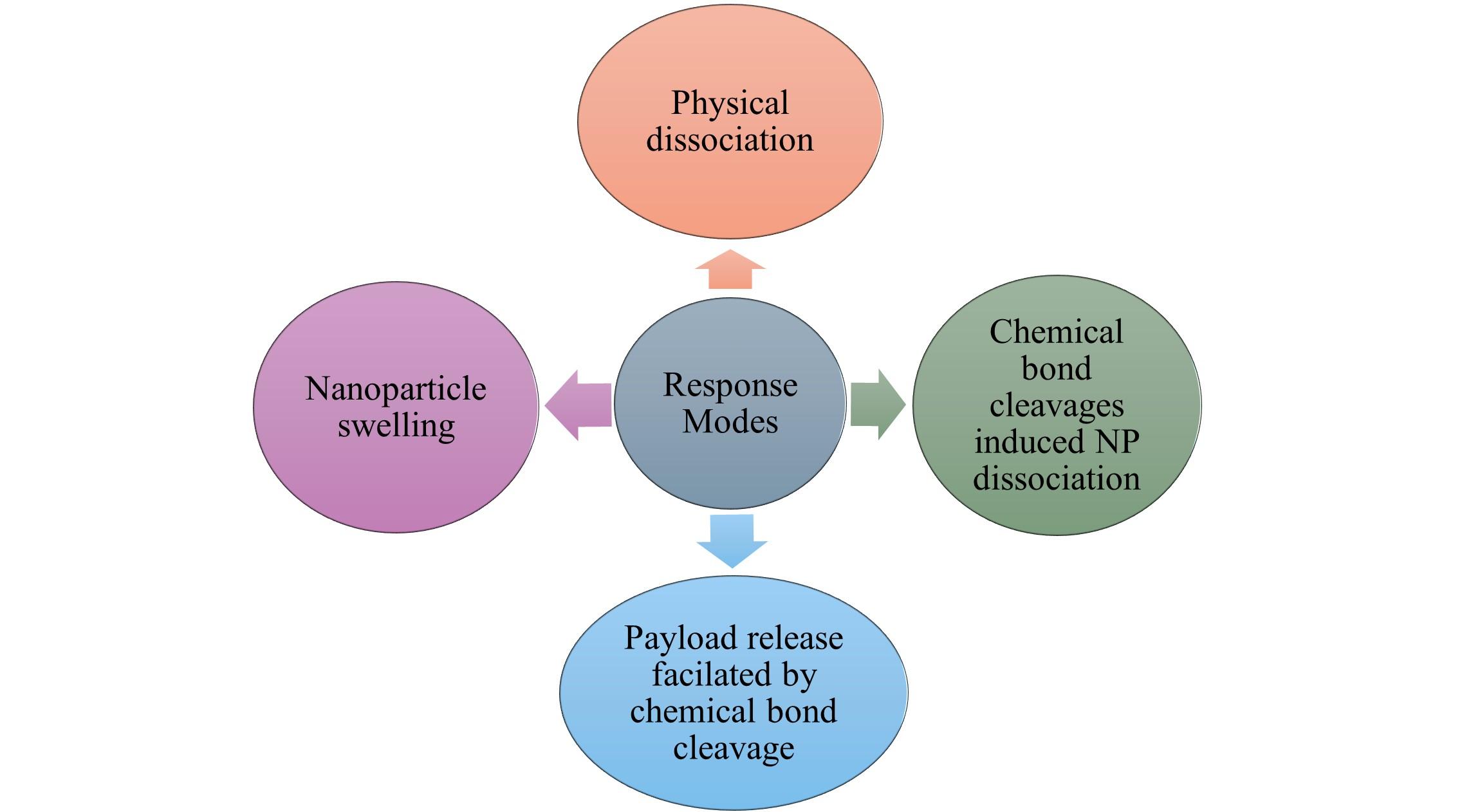
Physical dissociation
Physical dissociation refers to the process where nanoparticles undergo structural disintegration when exposed to a specific pH environment. This process is triggered by protonation or ionization of the hydrophobic core, leading to the loss of interactions that maintain the nanoparticle's stability. As a result, the nanoparticle breaks apart, allowing for the controlled release of its payload [35]. This property is particularly useful in the development of fluorescence probes and pH-sensitive drug delivery systems. By incorporating fluorescent dyes or therapeutic agents within the nanoparticle structure, they can function as switchable "ON-OFF" systems, responding dynamically to changes in pH. In an acidic environment, the protonation or ionization of hydrophobic segments weakens their interactions, leading to nanoparticle dissociation. This activates fluorescence or triggers drug release [36].
Physical and chemical changes in pH-sensitive polymers
At Neutral pH, the polymer remains structurally compact and stable, maintaining its integrity and preventing premature drug release.
At Acidic pH, functional groups such as carboxyl undergo ionization, leading to increased water absorption. This results in polymer expansion, which facilitates drug release.
At Basic pH, when exposed to an alkaline environment, polymers containing amine groups experience deprotonation. This can lead to polymer contraction or aggregation, potentially affecting the drug release profile [35].
The solubility of pH-sensitive polymers is highly dependent on their protonation and deprotonation states. These changes influence polymer behavior and drug delivery efficiency. Example: PAA remains insoluble at low pH due to the protonation of carboxyl groups, reducing charge repulsion. However, at higher pH levels, deprotonation increases charge repulsion, enhances solubility, and facilitates drug release [37].
Chemical bond cleavage that promotes payload release
In this method, hydrophobic regions of block copolymers are coupled with drug/fluorescent dyes via acid-labile chemical bonds to produce polymeric micelles [38]. When the polymeric micelles are in normal physiological states, the polymer gets stable and does not leak drugs or fluorescent agents. However, when the polymer reaches acidic sites (like tumors), the acid-sensitive chemical linkages will be hydrolyzed and release the drug/fluorescent dyes. The breaking chemical bond pH-sensitive nanosystems are made from polymers that have acid-labile chemical linkages [15].
Chemical bond cleavage leads to the dissociation of nanoparticles
This describes the process by which the hydrophobic and hydrophilic fragments cleave into nanoparticles, which leads to the dissolution of the nanoparticles. This method involves trapping drugs within self-forming nano-micelles by linking hydrophilic and hydrophobic parts of amphiphilic block copolymers using acid-sensitive chemical bonds. Under normal physiological pH conditions, the encapsulated drugs remain safely contained, and the micelles retain their stability without releasing their contents. Acid-labile bonds break when H+ penetrates the micelles in an acidic environment, causing the polymer to dissolve and the release of drugs. Hydrazone, imide, ester, ortho ester, acetal, and other acid-sensitive compounds are commonly used [39].
Nanoparticle swelling
The technique is known as "swelling mode," in which pH-sensitive nanoparticle micelles expand to release their payload [15, 31]. The concept behind the development of swelling polymeric micelles is the use of chemical bonds that are soluble in acid to connect hydrophobic segments to lengthy hydrophobic chains. Self-assembly in aqueous solutions is made possible by regulating the ratio of hydrophilic to hydrophobic blocks. H+ penetrates acidic environments, such as tumor tissues, where it helps to dissolve bonds, release hydrophobic fragments, increase solubility, enlarge the micelle volume, and leak payload [35].
pH-sensitive nanoparticle delivery for tumor targeting
Nanoparticles that respond to the acidic pH levels of certain tissue: Acidic pH levels ranging from 5.7 to 6.8 have been observed in human tumors [40]. The majority of tumors have inadequate blood supply and poor lymphatic drainage, which further contributes to the acidic tumor environment. pH-sensitive nanoparticles can aggregate in tumor tissue through improved permeability as well as retention effects, which facilitate both active and passive targeting [41]. The pH-dependent release of drugs through the use of polymers that vary their physical and chemical characteristics, such as swelling and solubility, according to local pH levels [42]. In figure 2, the delivery of the pH-sensitive nanoparticle at tumor cells is given.

Nanoparticles that respond to the basic environment: The gastrointestinal system keeps its pH levels consistent, ranging from the alkaline duodenum, ileum (pH 6.6-7.5) and cecum, rectum (6.0-7.0) for the stabilization of fluid to the acidic stomach lumen (pH 1-3) for digestion [43]. When a drug is encapsulated inside pH-sensitive nanoparticles, the nanoparticles can shield the drug from the stomach's acidic environment. These nanoparticles are designed to be stable in the acidic conditions of the stomach but will only release the drug when they reach a more neutral or slightly basic environment. Recent developments in nanotechnology involved surface-functionalized receptors for cellular targeting, transepithelial transport, and pH-sensitive methods for enhancing systemic absorption by increased stomach retention [44]. Making Nanoparticles with pH-dependent swelling is a commonly used strategy to accomplish organ-specific delivery of drugs. For example, when nanoparticles were carried with insulin, almost 90% of the insulin was released in its swollen form at pH 7.4 after two hours, but only 10% of the insulin was released in its collapsed condition at pH 1.2 [43].
pH-sensitive delivery of nanoparticles at the transversal region
At endosomal pH, Nanoparticles uptake protons, which raise the osmotic pressure within the endosomal segment, followed by rupture of the plasma membrane, which causes the nanoparticles to leak into the cytoplasm, and the pKa of the polymer can be adjusted for better endosomal access using copolymers that are composed of non-ionic and pH-sensitive monomers. For instance, it was feasible to modify the nanoparticle pH transfection efficiency by employing copolymers derived from monomers that had different pKa (such as dimethylaminoethyl methacrylate) [45]. Polymers with pH-sensitive properties in buffer endosomal compartments were grafted with additional functional regions for intracellular distribution. An example of amphiphilic and cationic triblock polymer was developed for siRNA transport and endosomal buffering, using monomethoxy polyethylene glycol (PEG), poly(ε-caprolactone), and poly (2-aminoethyl ethylene phosphate) [46].
Recent studies have focused on developing pH-sensitive polymeric nanocarriers that can effectively deliver antitumor agents. These smart nanocarriers are designed to permeate physiological barriers and release their payload in response to the acidic conditions found in tumor microenvironments. Such strategies aim to improve intracellular transport and target efficiency, addressing limitations associated with conventional chemotherapy delivery systems [47].
In the field of drug delivery, recent innovations in pH-sensitive nanoparticles have enhanced their precision and efficiency. Researchers have developed hybrid systems that combine polymers and lipids, enabling these nanoparticles to respond to acidic conditions found in specific body environments, such as the stomach or tumor tissues. These systems are particularly effective for delivering drugs like prednisolone, releasing the payload precisely at the desired site to minimize side effects and improve therapeutic outcomes. Additionally, advancements in polymer-based nanocomposites have led to dual-responsive systems that react to both acidic pH and redox conditions, which are common in tumor microenvironments. Such designs ensure a more targeted and efficient release of drugs, reducing the potential for off-target effects and improving treatment efficacy [48].
A promising development includes the introduction of cubosome liquid crystalline nanoparticles functionalized with pH-sensitive materials. These nanocarriers are capable of adapting to acidic environments, making them suitable for oral or topical drug delivery. This innovative approach has shown potential in addressing diseases where localized drug delivery is critical [49]. Furthermore, advancements in nanoparticle stabilization techniques, including surface coating and the creation of core-shell nanostructures, have been explored to enhance the stability and functionality of pH-sensitive nanoparticles in biological systems. These approaches aim to improve the therapeutic outcomes of nanoparticle-based drug delivery systems [49]. Various types of Smart drug delivery systems are listed in Table 3. The challenges of pH-sensitive nanoparticles in drug delivery are listed in Table 4.
Table 3. Comparison of pH-sensitive nanoparticles with other types of smart delivery systems.
Table 4. Challenges of pH-sensitive nanoparticles in drug delivery.
APPLICATION OF pH-SENSITIVE DRUG DELIVERY SYSTEM
Gastrointestinal drug delivery
The gastrointestinal system is one of the most often used modes of administration since it is simple to administer, has a controlled dose, and is very effective. Furthermore, a drug delivery system that is orally absorbed must be responsive to the pH changes in the gastrointestinal tract besides the ability to bypass the stomach acid. Oral drug-delivery systems use pH-sensitive polymers to maintain stability, prevent drug leakage into stomach juice, and release the medication into the intestines within 8-10 hours [15]. For example, Verapamil hydrochloride, a short-acting medication, is well-absorbed in the gastrointestinal tract, making it suitable for pH-sensitive formulations, particularly for treating diseases like gastritis and gastric cancer [66].
Drug delivery to tumor tissues and chemotherapy
The rapid growth of cancer cells in tumor sites results in an acidic environment, leading to increased glucose consumption, lactic acid accumulation, and restricted blood flow [67]. pH-sensitive nanoparticles have the potential to enhance the transportation of chemotherapeutic medicines to cancerous tumors, hence improving the efficacy of therapies. Molecular imaging, using target-specific probes, offers non-invasive viewing and statistical characterization of biological compounds in vivo, improving image-guided medication administration and surgical procedures [68, 69].
For example, nanomicelles monomethoxy polyethylene glycol-poly (lactic-co-glycolic acid) (mPEG-PLGA) with hydrophobic rare-earth nanoparticles. These nanoprobes, approximately 300 nanometers in size, are designed to respond to the acidic microenvironment of tumor tissues by breaking down and releasing chemotherapeutic drugs directly at the tumor site. Additionally, these nanoprobes enable imaging in the near-infrared II (NIR-II) range, providing high-resolution visualization for precise treatment monitoring and assisting in guiding surgical procedures [70].
Immunotherapy
The term "nano vaccine" refers to a type of pH-sensitive nanomaterial that may stimulate humorous or cellular immunity by transmitting antigens or immune activators for immunotherapy [15]. By using pH-sensitive compounds, different immunotherapy medications may be targeted more effectively, increasing their therapeutic impact and decreasing their harmful and adverse effects. For example, a Multifunctional oral nanoparticle delivery system loaded with anti-miR-301a (a microRNA inhibitor targeting miR-301a) aims to enhance targeting ability and therapeutic efficacy for inflammatory bowel disease [70].
pH-sensitive nano-delivery systems can conjugate or encapsulate fluorescent dyes, antigens, and small molecule medications, enabling medication distribution and disease site visualization [43].
Biosensors and gene carriers
pH-sensitive polymers are widely used to create insulin delivery devices for those who are diabetic. In reaction to blood glucose levels, the glucose-sensitive polymer technique can produce maintaining insulin, which can regulate the amount of insulin within normal levels. For example, Insulin-releasing beads were prepared by putting them in an aqueous solution using pH-sensitive polymers. Using pH-sensitive polymers as non-viral gene carriers is one of their most fascinating uses. The negative charge and enormous size of naked DNA under physiological environments make it extremely difficult to integrate into cells [71]. The transfer of genes intracellularly has garnered significant attention in recent times, owing to advancements in biotechnology and its potential medicinal applications. Gene transfection into vascular or cardiac cells has been explored using a variety of cationic polymers, such as linear or branched polyethylenimine, polyamidoamine dendrimer.
Advancements and strategies to enhance the targeting accuracy of pH-sensitive nanoparticles
The integration of both pH and light sensitivity into a single nanomaterial allows for more precise control of drug delivery, both through the body's natural pH changes and via external light sources, creating a more targeted and effective therapeutic strategy. As light-sensitive materials react to specific wavelengths of light, which is a powerful external trigger that can further activate or control the release of drugs. This dual-responsive behavior provides spatial and temporal control over the drug release. The system can be designed to respond to the acidic environment at the disease site, ensuring that the drug is released only when needed [72].
Temperature-sensitive nanoparticles, such as those made from poly (N-isopropyl acrylamide), undergo structural changes at specific temperatures, adjusting their solubility and permeability. This feature is useful for targeting tissues like tumors, which can be treated with hyperthermia. When combined with pH sensitivity, these nanoparticles can release drugs in tumor environments, ensuring targeted delivery. External heat or light sources can further refine drug release, enhancing precision.
For example, mesoporous silica nanoparticles (MSNs) modified for both temperature and pH sensitivity allow controlled drug release in acidic, heated tumor environments. This dual stimulus ensures that the drug is released only at the target site, minimizing side effects on healthy tissues.
Development of advanced materials
Recent advancements have led to the emergence of various materials that respond to pH changes, which are essential in the development of nanoparticles. Polymers like polyacrylic acid, polyhistidine, and polyglutamic acid have gained attention due to their ability to undergo structural and solubility alterations when exposed to different pH levels. These pH-sensitive polymers facilitate the controlled release of therapeutic agents, ensuring that drugs are only released in specific, acidic environments, such as those found in tumors or inflamed tissues. This precise targeting helps improve the efficacy of the treatment while reducing potential side effects [73].
Surface functionalization with targeting ligands
One of the most promising strategies to enhance the targeting accuracy of pH-sensitive nanoparticles is functionalization with targeting ligands. These ligands can recognize specific receptors on the surface of targeted cells, such as those found in tumors or inflamed tissues. This selective binding allows nanoparticles to accumulate at the site of interest, significantly improving the precision of drug delivery [74]. Recent advancements in drug delivery technology have resulted in the development of lectin-conjugated pH-sensitive mesoporous silica nanoparticles. These specially designed nanoparticles have the ability to recognize and bind to unique markers on the surfaces of specific cells, making them highly effective for targeting bone cancer cells. By ensuring that therapeutic agents are delivered directly to diseased tissues, this approach enhances treatment effectiveness while minimizing unintended effects on healthy cells. As a result, this strategy significantly improves the precision, safety, and overall efficiency of nanoparticle-based drug delivery systems [75].
The various types of characterization techniques for pH-sensitive nanoparticles are listed in Table 5.
Table 5. Characterization techniques of pH-sensitive nanoparticles.
RECENT RESEARCH AND PATENTS
LIMITATIONS AND FUTURE PROSPECTS
One of the problems confronted by pH-sensitive nanoparticles is variability in pH throughout the tumor growth and its dependency according to the type and stage of cancer. This inconsistency in tumor acidity can lead to unpredictable drug release, reducing treatment effectiveness. Additionally, these nanoparticles often suffer from poor targeting accuracy, leading to off-target effects and potential toxicity in healthy tissues. In these situations, receptor-mediated active targeting can help mitigate systemic toxicity by enhancing precision and specificity [93]. Another significant limitation lies in the biological barriers that hinder nanoparticle delivery. The body's mononuclear phagocyte system rapidly clears foreign particles from circulation, reducing the amount of nanoparticles that successfully reach tumor sites. Moreover, the tumor extracellular matrix presents a dense structural barrier that restricts deep penetration, making it difficult for nanoparticles to effectively distribute throughout the tumor mass. In addition, premature drug release triggered by pH variations in non-targeted areas can compromise the stability and effectiveness of these nanocarriers. Cancer cells often exhibit multidrug resistance (MDR), a mechanism that enables them to actively expel therapeutic compounds, decreasing treatment effectiveness. While pH-sensitive nanoparticles offer potential solutions, their ability to bypass MDR remains inadequate without additional modifications. Strategies such as altering nanoparticle composition and surface functionalization are being explored to improve intracellular drug retention. However, the materials used in these nanoparticles, typically synthetic polymers, raise concerns about their long-term accumulation and biodegradability, which could lead to unforeseen toxic effects over time. In oral drug delivery, maintaining bioavailability is challenging due to drug degradation in stomach acid, and large-scale production inconsistencies hinder clinical application. To overcome these limitations, researchers are developing multi-stimuli-responsive nanoparticles that combine pH sensitivity with other triggers like redox and enzymatic activity for better control and precision.
The future of pH-sensitive nanoparticles for drug delivery focuses on enhancing efficiency, stability, and large-scale applicability. Advanced biodegradable polymers will improve biocompatibility while reducing toxicity, ensuring safer therapeutic use. Multi-stimuli-responsive nanoparticles, which react to pH, temperature, and enzymes, will enable precise drug release. Targeted drug delivery through ligand-functionalized nanoparticles will minimize off-target effects and enhance treatment effectiveness.
Enhancing stability and shelf-life is crucial for clinical applications, ensuring nanoparticles remain effective under various conditions. Scaling Up Production- Creating pH-sensitive nanoparticles on a larger scale while maintaining consistency and quality is a significant challenge. So, focus on Mass Production Techniques and regulatory Standards. Additionally, personalized medicine will drive research into patient-specific nanoparticle designs, optimizing treatments based on individual profiles.
CONCLUSIONS
Several advanced methods have been used to develop pH-sensitive nanoparticles that can be used for therapeutic purposes (Figure 3). The fragments can engineer pH-sensitive nanoparticles and their response mechanisms, including chemical bond cleavage-induced dissociation, physical dissociation, and payload release via swelling nanoparticles. With the use of these techniques, the particles can precisely administer their medication in response to pH variations in the acidic regions. Furthermore, there is still much to learn about directing nanoparticles to certain areas. In vivo, many targeted systems exhibit little advantage over their non-targeted counterparts. The creation of standardized tests that could be used to compare newly produced particles to materials already in use would be beneficial for nanomedicine. To create a manual for improved material design, it is essential to integrate novel delivery methods with a deeper comprehension of the interactions between biological systems and nanoparticles.

AUTHOR CONTRIBUTIONS
The manuscript was drafted by PB and VJ. NN provided supervision throughout the manuscript preparation. Other authors contributed directly or indirectly to the revision and publication of this manuscript.
ACKNOWLEDGMENTS
The authors expressed gratitude to Mr. Jitender Joshi, President, Prof. (Dr.) Dharam Buddhi, Vice Chancellor of Uttaranchal Institute of Pharmaceutical Sciences, for their encouragement and guidance in the publication of this review work.
CONFLICTS OF INTEREST
There is no conflict of interest among the authors.
References
- [1]Ghaffar A, Yameen B, et al. pH-sensitive drug delivery systems. Metal Nanoparticles for Drug Delivery and Diagnostic Applications. 2020; 259–278.
- [2]Stuart MAC, Huck WTS, et al. Emerging applications of stimuli-responsive polymer materials. Nature Materials. 2010 9:2 2010; 9: 101–113.
- [3]Yoo JW, Giri N, et al. pH-sensitive Eudragit nanoparticles for mucosal drug delivery. Int J Pharm. 2011; 403: 262–267.
- [4]Deirram N, Zhang C, et al. pH-Responsive Polymer Nanoparticles for Drug Delivery. Macromol Rapid Commun. 2019; 40: 1800917.
- [5]Ijaz I, Gilani E, et al. Detail review on chemical, physical and green synthesis, classification, characterizations and applications of nanoparticles. Green Chem Lett Rev. 2020; 13: 59–81.
- [6]Shikuku R, Hasnat MA, et al. Chitosan-based pH-sensitive semi-interpenetrating network nanoparticles as a sustained release matrix for anticancer drug delivery. Carbohydrate Polymer Technologies and Applications. 2024; 7: 100515.
- [7]Li YY, Dong HQ, et al. Stimulus-responsive polymeric nanoparticles for biomedical applications. Science China Chemistry. 2010 53:3 2010; 53: 447–457.
- [8]Balamurali V, Pramodkuma TM, et al. pH Sensitive Drug Delivery Systems: A Review. American Journal of Drug Discovery and Development. 2010; 1: 24–48.
- [9]Shi Z, Li Q, Mei L. pH-Sensitive nanoscale materials as robust drug delivery systems for cancer therapy. Chinese Chemical Letters. 2020; 31: 1345–1356.
- [10]Selby LI, Cortez-Jugo CM, et al. Nanoescapology: progress toward understanding the endosomal escape of polymeric nanoparticles. Wiley Interdiscip Rev Nanomed Nanobiotechnol. 2017; 9: e1452.
- [11]Izumi H, Torigoe T, et al. Cellular pH regulators: potentially promising molecular targets for cancer chemotherapy. Cancer Treat Rev. 2003; 29: 541–549.
- [12]Liu J, Huang Y, et al. pH-Sensitive nano-systems for drug delivery in cancer therapy. Biotechnol Adv. 2014; 32: 693–710.
- [13]Wang X, Yang Y, et al. Fabrication of pH-Responsive Nanoparticles with an AIE Feature for Imaging Intracellular Drug Delivery. Biomacromolecules. 2016; 17: 2920–2929.
- [14]Such GK, Yan Y, et al. Interfacing Materials Science and Biology for Drug Carrier Design. Advanced Materials. 2015; 27: 2278–2297.
- [15]Mu Y, Gong L, et al. Advances in pH-responsive drug delivery systems. OpenNano .2021; 5: 100031.
- [16]Casey JR, Grinstein S, Orlowski J. Sensors and regulators of intracellular pH. Nat Rev Mol Cell Biol. 2010; 11: 50–61.
- [17]Ma X, Wang Y, et al. Ultra-pH-sensitive nanoprobe library with broad pH tunability and fluorescence emissions. J Am Chem Soc. 2014; 136: 11085–11092.
- [18]Liu L, Yao WD, et al. pH-Responsive carriers for oral drug delivery: challenges and opportunities of current platforms. Drug Deliv. 2017; 24: 569–581.
- [19]Zhuo S, Zhang F, et al. pH-Sensitive Biomaterials for Drug Delivery. Molecules 2020, Vol 25, Page 5649 2020; 25: 5649.
- [20]Shi Z, Li Q, et al. pH-Sensitive nanoscale materials as robust drug delivery systems for cancer therapy. Chinese Chemical Letters. 2020; 31: 1345–1356.
- [21]Lin Z, Zhu H, et al. Direct introduction of cationic and anionic lipids to create pH-sensitive charge-reversible liposomes with optimized pharmacokinetics and antitumor effects. Journal of Nanoparticle Research. 2024; 26: 1–14.
- [22]Dilshener D, Parsons DF, et al. pH-sensitive spontaneous decay of functionalized carbon dots in solutions. The Journal of Chemical Physics.2024; 160; 212103
- [23]Sakurai Y, Watanabe H, et al. pH-Responsive Lipid Nanoparticles Achieve Efficient mRNA Transfection in Brain Capillary Endothelial Cells. Pharmaceutics. 2022; 14: 1560.
- [24]Jha A, Rama A, et al. Temperature and pH-responsive nanogels as intelligent drug delivery systems: A comprehensive review. J Appl Pharm Sci. 2021; 11: 001–016.
- [25]Torchilin V, et al. Multifunctional and stimuli-sensitive pharmaceutical nanocarriers. European Journal of Pharmaceutics and Biopharmaceutics. 2009; 71: 431–444.
- [26]Kumar N, Singh S, et al. Single-, Dual-, and Multi-Stimuli-Responsive Nanogels for Biomedical Applications. Gels. 2024, Vol 10, Page 61 2024; 10: 61.
- [27]Malikmammadov E, Hasirci N. Dual- and Multistimuli-Responsive Polymers for Biomedical Applications. Smart Polymers and Their Applications. 2019; 255–278.
- [28][Sethuraman V, Janakiraman K, et al. Recent Progress in Stimuli-Responsive Intelligent Nano Scale Drug Delivery Systems: A Special Focus Towards pH-Sensitive Systems. Curr Drug Targets. 2021; 22: 947–966.
- [29]Singh J, Nayak P. pH-responsive polymers for drug delivery: Trends and opportunities. Journal of Polymer Science. 2023; 61: 2828–2850.
- [30]Ma X, Wang Y, et al. Ultra-pH-sensitive nanoprobe library with broad pH tunability and fluorescence emissions. J Am Chem Soc. 2014; 136: 11085–11092.
- [31]Wang Z, Deng X, et al. Mechanisms of drug release in pH-sensitive micelles for tumour targeted drug delivery system: A review. Int J Pharm. 2018; 535: 253–260.
- [32]Gupta P, Vermani K, et al. Hydrogels: from controlled release to pH-responsive drug delivery. Drug Discov Today. 2002; 7: 569–579.
- [33]Lu Y, Sun W, et al. Stimuli-responsive nanomaterials for therapeutic protein delivery. Journal of Controlled Release. 2014; 194: 1–19.
- [34]Li Y, Wang Z, et al. Non-covalent interactions in controlling pH-responsive behaviors of self-assembled nanosystems. Polym Chem. 2016; 7: 5949–5956.
- [35]Mu Y, Gong L, et al. Advances in pH-responsive drug delivery systems. OpenNano. 2021; 5: 100031.
- [36]Zhang G, Yue K, et al. Self-assembly and disassembly mechanisms of biomimetic peptides: Molecular dynamics simulation and experimental measurement. Int J Biol Macromol. 2022; 209: 785–793.
- [37]Chatterjee S, Hui PC leung. Review of Stimuli-Responsive Polymers in Drug Delivery and Textile Application. Molecules. 2019; 24:14; 2547.
- [38]Li Y, Zhao T, et al. Molecular basis of cooperativity in pH-triggered supramolecular self-assembly. Nat Commun7. 2016; 13214
- [39]Thambi T, Deepagan VG, et al. Synthesis and physicochemical characterization of amphiphilic block copolymers bearing acid-sensitive orthoester linkage as the drug carrier. Polymer (Guildf). 2011; 52: 4753–4759.
- [40]Vaupel P. Tumor microenvironmental physiology and its implications for radiation oncology. Semin Radiat Oncol. 2004; 14: 198–206.
- [41]Shinn J, Kwon N, et al. Smart pH-responsive nanomedicines for disease therapy. J Pharm Investig. 2022; 52: 427.
- [42]Gao W, Chan JM, et al. pH-responsive Nanoparticles for Drug Delivery. Mol Pharm. 2010; 7: 1913.
- [43]Gao W, Chan JM, et al. PH-responsive nanoparticles for drug delivery. Mol Pharm. 2010; 7: 1913–1920.
- [44]Yamanaka YJ, Leong KW. Engineering strategies to enhance nanoparticle-mediated oral delivery. J Biomater Sci Polym Ed. 2008; 19: 1549–1570.
- [45]You JO, Auguste DT. Nanocarrier cross-linking density and pH sensitivity regulate intracellular gene transfer. Nano Lett. 2009; 9: 4467–4473.
- [46]Sun TM, Du JZ, et al. Self-assembled biodegradable micellar nanoparticles of amphiphilic and cationic block copolymer for siRNA delivery. Biomaterials. 2008; 29: 4348–4355.
- [47]Qin J, Zhu Y, et al. pH-sensitive polymeric nanocarriers for antitumor biotherapeutic molecules targeting delivery. Bio-Design and Manufacturing. 2021 4:3 2021; 4: 612–626.
- [48]Gaddimath S, Payamalle S, et al. Recent Advances in pH and Redox Responsive Polymer Nanocomposites for Cancer Therapy. Journal of Composites Science. 2024, Vol 8, Page 28 2024; 8: 28.
- [49]Ezike TC, Okpala US, et al. Advances in drug delivery systems, challenges and future directions. Heliyon. 2023; 9: e17488.
- [50]Huh KM, Kang HC, et al. pH-sensitive polymers for drug delivery. Macromol Res. 2012; 20: 224–233.
- [51]Kanamala M, Wilson WR, et al. Mechanisms and biomaterials in pH-responsive tumour targeted drug delivery: A review. Biomaterials. 2016; 85: 152–167.
- [52]Bi H, Xue J, et al. Current developments in drug delivery with thermosensitive liposomes. Asian J Pharm Sci. 2019; 14: 365–379.
- [53]Priya S, Jain KK, et al. Revolutionizing rheumatoid arthritis treatment with emerging cutaneous drug delivery systems: overcoming the challenges and paving the way forward. Nanoscale. 2024; 17: 65–87.
- [54]Kapalatiya H, Madav Y, et al. Enzyme-responsive smart nanocarriers for targeted chemotherapy: an overview. Drug Deliv Transl Res. 2022; 12: 1293–1305.
- [55]Li M, Zhao G, et al. Enzyme-Responsive Nanoparticles for Anti-tumor Drug Delivery. Front Chem. 2020; 8: 556896.
- [56]Selim MM, El-Safty S, et al. A review of magnetic nanoparticles used in nanomedicine. APL Mater. 2024; 010601
- [57]Zhang H, Liu XL, et al. Magnetic nanoparticles based cancer therapy: current status and applications. Sci China Life Sci. 2018; 61: 400–414.
- [58]Samanta D, Meiser JL, et al. Polypyrrole nanoparticles for tunable, pH-sensitive and sustained drug release. Nanoscale. 2015; 7: 9497–9504.
- [59]Liu L, Yao WD, et al. pH-Responsive carriers for oral drug delivery: challenges and opportunities of current platforms. Drug Deliv. 2017; 24: 569–581.
- [60]Yaşayan G, Alarcin E, et al. Biocompatibility and toxicity challenges of nanomaterials. Functionalized Nanomaterials for Cancer Research. 2024; 603–631.
- [61]Li X, Wang L, et al. Biocompatibility and Toxicity of Nanoparticles and Nanotubes. J Nanomater. 2012; 2012: 548389.
- [62]Missaoui WN, Arnold RD, et al. Toxicological status of nanoparticles: What we know and what we don't know. Chem Biol Interact. 2018; 1;295:1-12.
- [63]Fu PP, Xia Q, et al. Mechanisms of nanotoxicity: generation of reactive oxygen species. 2014; 22(1):64-75.
- [64]Hoffman AS. Hydrogels for biomedical applications. Adv Drug Deliv Rev. 2012; 64: 18–23.
- [65]Huh KM, Kang HC, et al. pH-sensitive polymers for drug delivery. Macromol Res. 2012; 20: 224–233.
- [66]Garg T, Kumar A, et al. Gastroretentive Drug Delivery Systems for Therapeutic Management of Peptic Ulcer. Critical Reviews & trade; in Therapeutic Drug Carrier Systems. 2014; 31: 531–557.
- [67]Tang H, Zhao W, et al. Recent Development of pH-Responsive Polymers for Cancer Nanomedicine. Molecules. 2019, Vol 24, Page 4 2018; 24: 4.
- [68]Nayak AK, Hasnain MS, et al. Drug delivery using interpenetrating polymeric networks of natural polymers: A recent update. J Drug Deliv Sci Technol. 2021; 66: 102915.
- [69]Xu L, Bai E, et al. pH-Responsive Hydrogel as a Potential Oral Delivery System of Baicalin for Prolonging Gastroprotective Activity. Pharmaceutics. 2023, Vol 15, Page 257 2023; 15: 257.
- [70]Feng M, Wang Y, et al. Degradable pH-responsive NIR-II imaging probes based on a polymer-lanthanide composite for chemotherapy. Dalton Transactions. 2020; 49: 9444-9453.
- [71]Reyes-Ortega F. pH-responsive polymers: properties, synthesis and applications. Smart Polymers and their Applications. 2014; 45–92.
- [72]Shin Y, Husni P, et al. Recent Advances in pH- or/and Photo-Responsive Nanovehicles. Pharmaceutics. 2021; 13: 725.
- [73]Kocak G, Tuncer C, et al. pH-Responsive polymers. Polym Chem. 2016; 8: 144–176.
- [74]Emerich DF, Thanos CG. Targeted nanoparticle-based drug delivery and diagnosis. J Drug Target. 2007; 15: 163–183.
- [75]Martinez-Carmona M, Lozano D, et al. Lectin-conjugated pH-responsive mesoporous silica nanoparticles for targeted bone cancer treatment. Acta Biomater. 2021; 65: 393–404.
- [76]Khan Y, Sadia H, et al. Classification, Synthetic, and Characterization Approaches to Nanoparticles, and Their Applications in Various Fields of Nanotechnology: A Review. Catalysts. 2022; 12: 1386.
- [77]Griset AP, Walpole J, et al. Expansile nanoparticles: Synthesis, characterization, and in vivo efficacy of an acid-responsive polymeric drug delivery system. J Am Chem Soc. 2009; 131: 2469–2471.
- [78]Belman Flores, et al. Synthesis and characterization of pH sensitive hydrogel nanoparticles based on poly (N-isopropyl acrylamide-co-methacrylic acid). Journal of Materials Science: Materials in Medicine. 2020; 31: 61.
- [79]Rehman S, Jamil QA, et al. Preparation and Evaluation of pH-Sensitive Chitosan/Alginate Nanohybrid Mucoadhesive Hydrogel Beads: An Effective Approach to a Gastro-Retentive Drug Delivery System. Pharmaceutics. 2024; 16: 1451.
- [80]Lopez-Vidal L, Parodi P, et al. Formulation and optimization of pH-sensitive nanocrystals for improved oral delivery. Drug Deliv Transl Res. 2024; 14: 1301–1318.
- [81]Alejo T, Toro-Córdova A, et al. Comprehensive Optimization of a Freeze-Drying Process Achieving Enhanced Long-Term Stability and In Vivo Performance of Lyophilized mRNA-LNPs. Int J Mol Sci. 2024; 25: 10603.
- [82]Yannan D, et al. Prioritizing Formulation Strategies for Temperature-Sensitive Biotherapeutics. Journal of Controlled Release. 2017; 249: 63-73.
- [83]Unsoy G, Khodadust R, et al. Synthesis of Doxorubicin loaded magnetic chitosan nanoparticles for pH responsive targeted drug delivery. European Journal of Pharmaceutical Sciences. 2014; 62: 243–250.
- [84]Chang D, Gao Y, et al. Polydopamine-based surface modification of mesoporous silica nanoparticles as pH-sensitive drug delivery vehicles for cancer therapy. J Colloid Interface Sci. 2016; 463: 279–287.
- [85]Rao Z, Ge H, et al. Carboxymethyl cellulose modified graphene oxide as pH-sensitive drug delivery system. Int J Biol Macromol. 2018; 107: 1184–1192.
- [86]Zhao T, Maniglio D, et al. Development of pH-sensitive self-nanoemulsifying drug delivery systems for acid-labile lipophilic drugs. Chem Phys Lipids. 2016; 196: 81–88.
- [87]Lin YH, Chang CH, et al. Development of pH-responsive chitosan/heparin nanoparticles for stomach-specific anti-Helicobacter pylori therapy. Biomaterials. 2009; 30: 3332–3342.
- [88]Pohlit H, Bellinghausen I, et al. Sting activating nano vaccine for immunotherapy. Biomacromolecules. 2017; 16: 3103–3111.
- [89]Adriamycin prodrug antitumor preparation. 2023.
- [90]Lignin-based pH-responsive magnetic nano-drug carrier and preparation method and application thereof. 2022.
- [91]Schiff base copolymer coated mesoporous silica drug-loaded nanoparticle with pH responsiveness and preparation method and application thereof. 2020.
- [92]Oct. Ph-responsive silica metal organic framework nanoparticles for delivery of bioactive molecules. 2022.
- [93]Alsawaftah NM, Awad NS, et al. pH-Responsive Nanocarriers in Cancer Therapy. Polymers. 2022 ; 14: 936.